

An accomplished professor’s career-long quest to understand peripheral nerve blocks has introduced new technologies and called into question decades of anesthesiology doctrine, in turn opening new frontiers in pain medicine.

Over the past two years, Andre Boezaart, MD, PhD, in collaboration with other faculty members in the Department of Anesthesiology’s Division of Acute and Perioperative Pain Medicine and researchers in Spain, has published a series of manuscripts casting doubt on the prevailing wisdom that intraneural injections necessarily cause neurological injury.
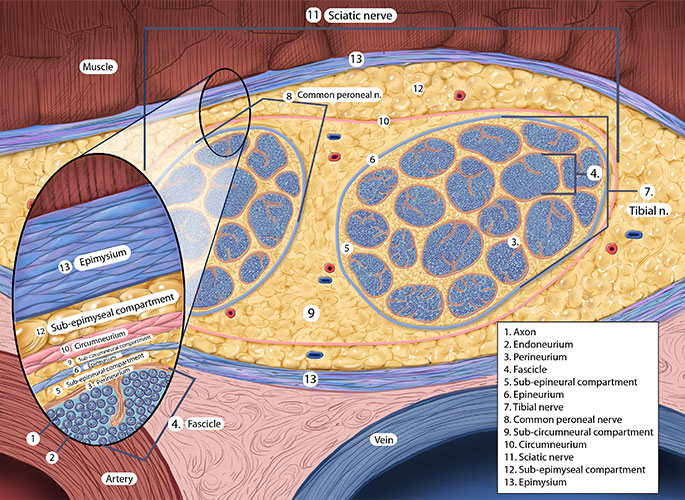
Their work has argued that the scientific community’s understanding was based on studies in animals, which do not correlate with human anatomy, especially microanatomy, and relied on markers such as India ink that spread from the compartments they are injected into.
The group further published research showing that it may be impossible to inject directly into a nerve fascicle, the functional unit of a nerve. Most recently, a manuscript in press describes a previously unknown layer of collagen that surrounds the nerve. The implication is that the electrical current in that layer is a direct current that helps with tissue regeneration, enabling electricity to be used as a therapeutic, said Dr. Boezaart, who retired from the department after 12 years in June.
The origin of his work dates to 1975 when Dr. Boezaart, conscripted into the South African Defense Force, was sent to Angola for Operation Savannah. Working as an anesthesiologist, Dr. Boezaart encountered serious hand injuries that required numerous follow-up surgeries.
Physicians were performing debridements of the hands every day and were using regional blocks, which were time consuming. They decided instead to insert catheters on the nerves in the axilla of the arm that could remain in place when patients were discharged and be used for repeated surgery and pain management. The result was the first-recorded continuous nerve block and first continuous nerve blocks used for transport of wounded soldiers, which eliminated the need for narcotics.
Years later, while Dr. Boezaart was working as an anesthesiologist at the Cape Shoulder Institute in Cape Town, South Africa, the team was unsuccessful at continuous blocks for shoulder surgery. Dr. Boezaart was prompted by a surgeon to consider what would happen if a nerve stimulator was placed on a catheter for a continuous block, as it is on a single-shot block.
“It changed the game,” Dr. Boezaart said. “Our continuous blocks became very successful and we started to send patients home in the early 1990s with continuous nerve blocks after major shoulder and other major surgeries without the need for opioids.”
Since then, he has performed about 25,000 such continuous blocks with stimulating catheters and also has a patent for the device. But the reason this technique worked so much better than other techniques, including ultrasound, remained mostly unexplained.

In 2015, Miguel Reina, MD, PhD, who works in an anesthesiology department that comprises six university hospitals in Madrid, published a book on microanatomy that collected 25 years of his work with electron and regular microscopy of nerves. It took a pair of outside eyes to see that the pictures opened the door for a new understanding of the nerve and explained why continuous nerve blocks worked or failed.
“I saw in the book what even Professor Reina didn’t know was there. He also admitted that every time he looked at his photographs he saw something new in them,” Dr. Boezaart said.
The book and the research that the two embarked on together revealed that the block works because the catheters were placed deep to a specific membrane, the circumneurium, that cannot be seen with ultrasound.
Dr. Boezaart ultimately traveled to Madrid in 2017 for a visiting professorship with Olga “Kiki” Nin, MD, Assistant Professor of Anesthesiology at UF.
One of the manuscripts that has been published through this collaboration, a PhD project with Anna Server, MD, PhD, described the revelation that animal data cannot be extrapolated to humans because of the vast microanatomical differences between the two. Another found that heparinized blood solution is superior to other markers such as India ink and methylene blue to identify the compartments surrounding nerves.
This work is important because a better understanding of nerve microanatomy helps to assess the effectiveness and risks of blocks.
This understanding was advanced when Paul Bigeleisen, MD, PhD, Professor of Anesthesiology at the University of Maryland and now a member of Dr. Boezaart’s research team, published work showing that nerve puncture or even intraneural injection does not inevitably cause neurologic injury.
The question then became whether injecting into the nerve fascicle caused injury.
Dr. Boezaart and his team completed a study in which they intraneurally injected heparinized blood solution with ultrasound guidance into fresh cadavers. After about 600 injections, they found that they could not inject into the relatively solid endoneurium of the fascicle, but only into fat-containing compartments outside the fascicles and among the cells of the perineurium, the membrane that surrounds the fascicles. The work was published in Anesthesiology.
“We thus debunked two principles held by the science of 100 years here, namely that it was valid to extrapolate animal experiments to humans and that injection into fascicles was possible,” Dr. Boezaart said.
The implication is that nerve injuries are more likely to be caused by other factors, such as traction, which is external pressure exerted on the nerve, or even genetic factors, rather than intraneural injections, Dr. Boezaart said. This is crucial because many anesthesiologists have experienced repercussions as severe as losing their licenses because of injuries blamed on blocks.
The findings have been met with resistance in the scientific community, in part because many fear that intraneural injections have the potential to cause patients harm and should not be normalized. Although Dr. Boezaart’s team in no way wants to normalize intraneural injection, they continue to seek the truth behind nerve injury.
Dr. Boezaart and his team thus took the work further, showing through the examination of nerves from fresh cadavers that there is a layer of collagen around the nerve, an internal epineurium, which forms a second direct current of electricity responsible for tissue regeneration. (The other electrical current is the alternating current of the action potential of the nerve axons). Because this second electrical current flows in only one direction, it can be used or enhanced for recovery of injured nerves or pain management, analogous to acupuncture. The results of this study are detailed in a manuscript under review in Clinical Anatomy.
While spinal cord stimulators use a similar concept and are well-established, the use of a neuromodulator for peripheral nerves is in its infancy, and the team is working on different electrical wave forms to further unravel it, Dr. Boezaart said.
The next step in the research, expected to begin this summer under the direction of Dr. Nin, is measuring the electrical current of the collagen layer with high-frequency ultrasound and microneedles. The team is also studying the treatment of diabetic peripheral neuropathy with neuromodulation under the leadership of Linda Le-Wendling, MD, Associate Professor of Anesthesiology, and Ajay Antony, MD, Assistant Professor of Anesthesiology, both at UF.
While it could take another decade for research to further substantiate the work, Dr. Boezaart, who is director of the Alon P. Winnie Research Institute, headquartered in South Africa, said he intends to continue reaching for better understanding, with the ultimate goal of mitigating human pain and suffering.